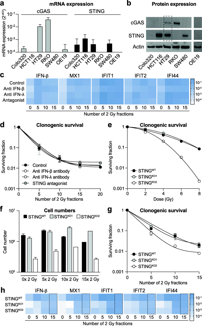
Fig. 4.
ISG induction upon fractionated irradiation occurs independent of STING, IFN-β or IFN-λ. Interferon stimulated genes (ISGs) can be induced independent of the interferons known to mediate this response, or the upstream regulator protein STING. a mRNA expression levels of cGAS (grey bars) and STING (black bars) in different cancer cell lines. b Western blots showing protein expression of cGAS and STING in different cancer cell lines. Actin staining was used as loading control. The dotted box shows the only cell line, i.e. HT29, in which both cGAS and STING protein expression could be detected. c Heat map of mRNA expression of different ISGs and IFN-β in HT29 cells treated with fractionated irradiation in the presence or absence of either anti IFN-β antibody, anti IFN-λ antibody or a STING antagonist. No significant changes were observed in the presence of any of the treatments as compared to irradiation alone (n = 3). d Clonogenic survival of HT29 during fractionated irradiation in the presence or absence of either anti IFN-β antibody, anti IFN-λ antibody or a STING antagonist. No significant changes in surviving fractions were observed in the presence of any of the latter treatments as compared to irradiation alone (n = 3). e Clonogenic survival of HT29 wild-type cells and two HT29 STING knockout cells in response to single dose irradiation. STINGKO2 shows higher radiosensitivity as compared to wild type cells. f Cell numbers of HT29 wild-type cells and two HT29 STING knockout cells during fractionated irradiation. While STINGKO2 displayed slower growth already at base-line, fractionated irradiation did not affect growth of knockout cells compared to wild type cells. g Clonogenic survival of HT29 wild-type cells and two HT29 STING knockout cells during fractionated irradiation. STINGKO2 shows higher radiosensitivity as compared to wild type cells. h) Heat map of mRNA expression of different ISGs and IFN-β in HT29 wild-type cells and two HT29 STING knockout cells during fractionated irradiation (n = 2). At baseline (0 × 2 Gy) both knockout cell lines show lower expression of all genes analyzed as compared to wild-type cells. At the end of the treatment period (15 × 2 Gy, dotted box) no more difference in expression levels is observed in wild-type vs. knockout cells for any of the genes analyzed

