Fig. 4.
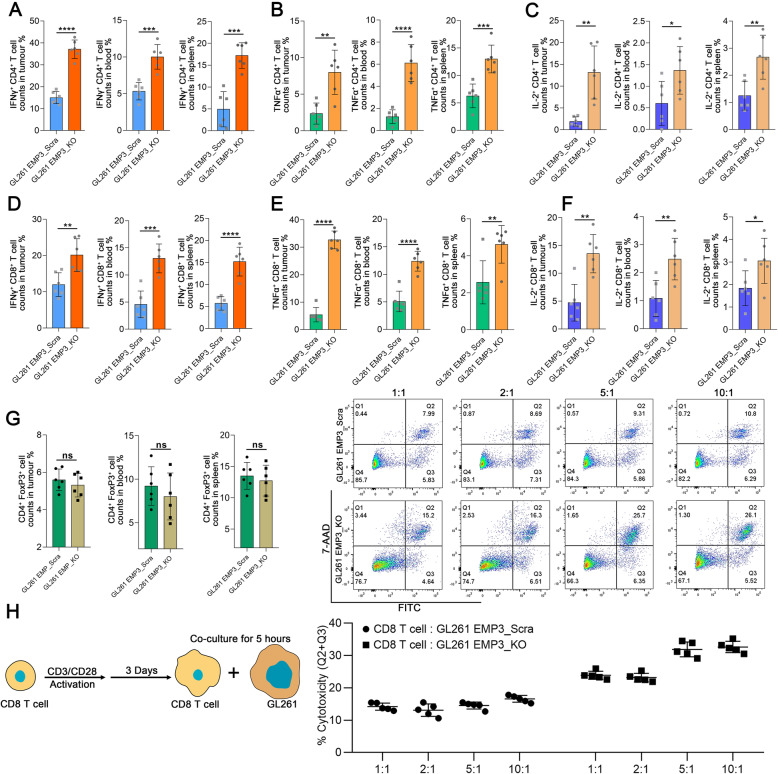
EMP3 drives T cell exclusion from the GBM microenvironment. a-c Percentages of IFN-γ+/TNF-α+/IL-2+ CD4+ T cells in mouse tumours, blood, and spleens from the EMP3_KO and EMP3_Scra GL261 groups. Student’s t-test was performed. d-f Percentages of IFN-γ+/TNF-α+/IL-2+CD8+ T cells in mouse tumours, blood, and spleens from the EMP3_KO and EMP3_Scra GL261 groups. Data were analysed using Student’s t-test. g Percentages of FoxP3+ CD4+ T cells in mouse tumours, blood, and spleens from the EMP3_KO and EMP3_Scra GL261 groups. Student’s t-test was performed.h CD8+ T cells were isolated from splenocytes from C57BL/6 mice and activated with anti-CD3/CD28 Dynabeads for 3 days. Activated CD8+ T cells were co-cultured with EMP3_KO or EMP3_Scra GL261 cells at ratios of 1:1, 2:1, 5:1, and 10:1. Cell apoptosis analysis was conducted with the Annexin V-FITC Apoptosis Detection Kit (BD Biosciences, USA) according to the manufacturer’s instructions. Student’s t-test was performed. The mean ± S.D. is shown. Ns: nonsignificant, *p < 0.05, **p < 0.01, and ***p < 0.001

