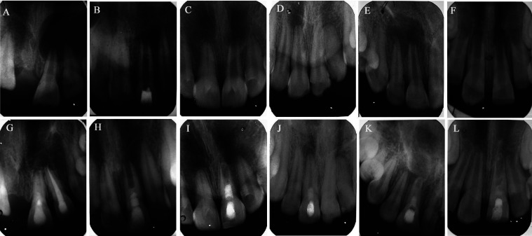
Figure 4.
Pre-operative radiographs with periapical lesion and immature apex (A,B and C for A-PRF and D,E and F for PRF) and final follow-up respective radiographs (G,H and I for A-PRF and J, K and L for PRF). (G) #21, A-PRF treated, 20-year-old male with a 16-month follow-up shows periapical healing with increased root thickening and root maturation. (H) #21, A-PRF treated, 17-year-old male with 17-months follow-up shows periapical healing with no significant continuation of root development. (I) #21, A-PRF treated, 12-year-old male, a 15-months follow-up reveals periapical healing with closed root apex (J) #11, PRF treated, 12-years-old male with 12 months follow-up, minimal periapical healing with thickening of canal walls and continued root maturation (K) #11, PRF treated, 17-year-old, male with 12-months follow-up, periapical healing and root apex closed (L) #21, PRF-treated, 8-year-old female, 12 months follow-up, periapical healing with root apex closed.

