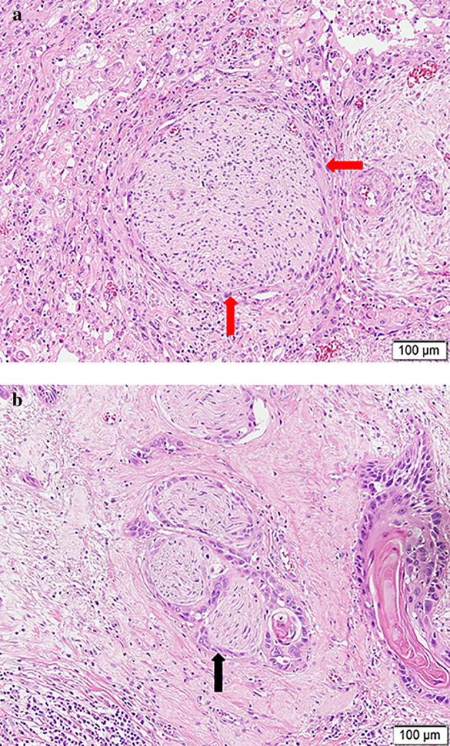
FIGURE 1.

The presence of perineural invasion in an esophageal squamous cell carcinoma specimen stained with hematoxylin and eosin. (a) The nerve fiber was partially surrounded by tumor cells (red arrow). (b) Tumor cells embedded in the perineurium (black arrow)
