FIGURE 2.

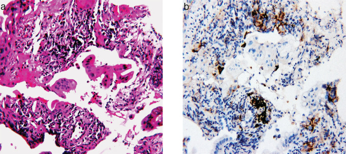
Microscopic examination. (a) Hematoxylin and eosin staining was consistent with adenocarcinoma. (b) Programmed death ligand 1 (PD‐L1) immunohistochemical staining of lung adenocarcinoma specimens with a tumor proportion score of 1%–49%

Microscopic examination. (a) Hematoxylin and eosin staining was consistent with adenocarcinoma. (b) Programmed death ligand 1 (PD‐L1) immunohistochemical staining of lung adenocarcinoma specimens with a tumor proportion score of 1%–49%