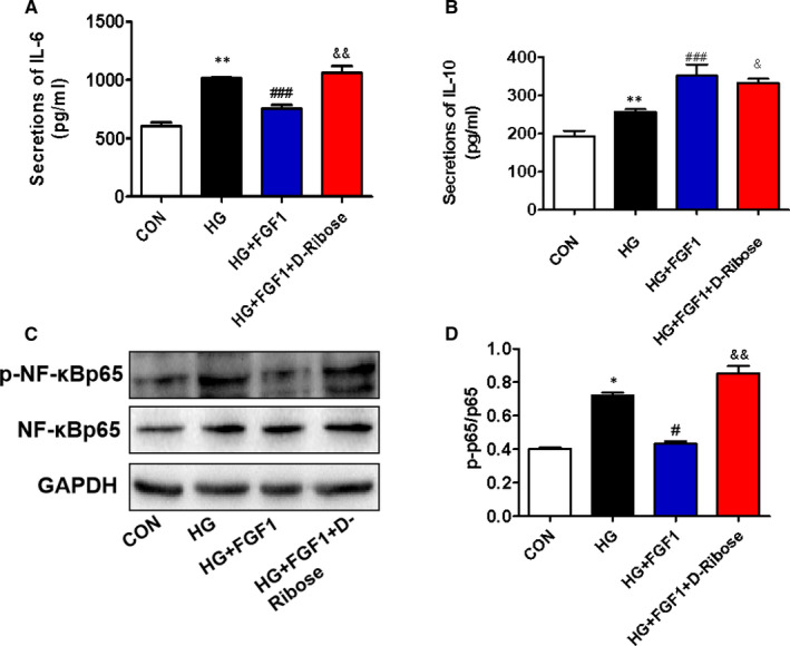
FIGURE 6.

D‐ribose blocks the role of FGF1 on high glucose–associated excessive inflammation in AML12 cells. (A and B) The enzyme‐linked immunosorbent assay (ELISA) shows the levels of pro‐inflammatory cytokines (IL‐6) and anti‐inflammatory cytokines (IL‐10) in AML12 cells from different groups, n = 3. C, The Western blotting and quantitative analyses of p‐NF‐κBp65 and NF‐κBp65 in AML‐12 cells from different groups, n = 3. *P < .05, **P < .01 vs CON group; # P < .05, ### P < .001 vs HG group; & P < .05, && P < .01 vs HG+FGF1 group
