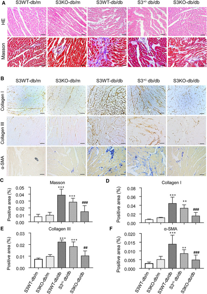
FIGURE 2.

Smad3 deficiency prevents db/db mice from cardiac fibrosis. A, Representative images of H&E and Masson's trichrome staining. B, Representative immunohistochemical images of collagen I, collagen III and α‐SMA. C, Quantitative analysis of Mason's trichrome staining. D‐F, Quantitative analysis of collagen I, collagen III and α‐SMA immunohistochemical staining. Values are expressed as mean ± SE for group of eight mice. **P <.01 and ***P <.001 compared with Smad3(S3) WT‐db/m group; ## P <.01 and ### P <.001 compared with Smad3 WT‐db/db and Smad3+/−db/db groups. Scale bar = 20 µm
