
Figure 1.
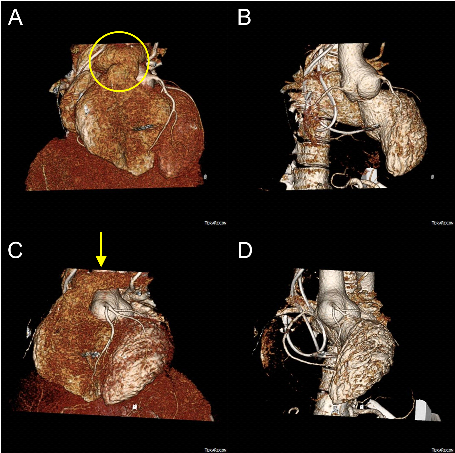
ECG-Gated CT Angiography with 3D postprocessing is shown for images pre-surgery. 3D volume rendered CTA images from multiple orientations are shown in A)-D). The overlying pulmonary artery is shown with a yellow oval (A) and the location of the incision of the left pulmonary artery is shown with a yellow arrow (C). Reconstructed images with the right ventricle removed are shown in B) and D) to view the aortic root aneurysm.