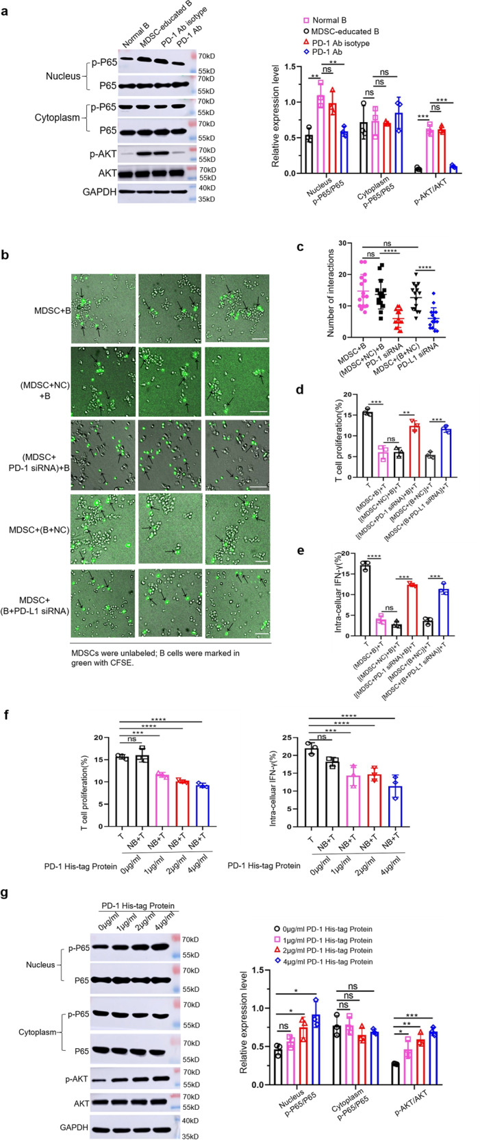
Fig. 3. Tumor-derived MDSCs activate the PI3K/AKT/NF-κB signaling pathway in B cells through PD-1/PD-L1 axis.

a Tumor-derived MDSCs were cocultured with normal B cells in the presence or absence of anti-PD-1 Ab and the protein levels of AKT, p-AKT, P65 and p-P65 were measured by western blot (n = 3). b, c MDSCs transfected with PD-1 siRNA, or B cells transfected with PD-L1 siRNA were cocultured with either B cells or MDSCs. For each group, we selected three different fields of view to analyze the mutual contacts between MDSCs with B cells by live cell imaging (n = 3). Scale bar: 50 µm. NC negative control. d B cells were isolated from the above five groups and co-incubated with T cells for 48 h. T cell proliferation and IFN-γ secretion (e) were measured (n = 3). f Normal B cells were stimulated with different concentrations of recombinant PD-1 His-tagged protein (R&D Systems, USA) and cultured with T cells for 48 h. T cell proliferation and IFN-γ secretion (n = 3), as well as protein levels of AKT, p-AKT, P65, and p-P65 were measured (g). (In all experiments, Bar graphs and plots represent or include mean ± SD, respectively. ns no statistically significant, *p < 0.05, **p < 0.01, ***p < 0.001, ****p < 0.0001).
