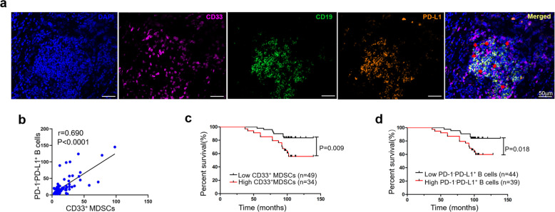
Fig. 6. PD-1−PD-L1+ Bregs and MDSCs are colocalized in tumor tissues and associated with poor prognosis in breast cancer+.
a Multiplex fluorescent immunohistochemistry staining of breast cancer tissue showing CD33 (purple), CD19 (green), and PD-L1 (orange) in close proximity (red arrows). Original images were taken at ×20 magnification. Scale bar: 50 µm. b The number of PD-1−PD-L1+ Bregs (n = 83) was correlated with the number of CD33+ MDSCs (n = 83). Kaplan–Meier analysis graph showing that MDSCs (c) and PD-1−PD-L1+ Bregs (d) were associated with the overall survival of patients (n = 83) with breast cancer.

