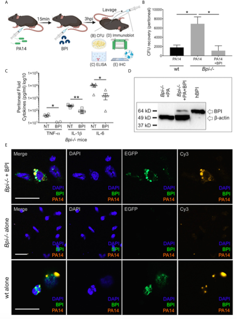
Figure 4.
Exogenous BPI mediates P. aeruginosa uptake in Bpi-/- mice in vivo. (A) Mice were administered 10μg BPI 15mins following PA14 infection (3x10⁶ CFU) via intraperitoneal route. Peritoneal lavage was performed 3hpi for PA14 colony count, ELISA, immunoblot, and IHC. Administration of neutrophil-purified human BPI (hBPI, 10μg) into the peritoneal cavity of mice infected with PA14 (3x10⁶ CFUs) lowers (B) bacterial colony forming units (CFU) recovered from peritoneal fluid of Bpi-/- mice (n=4), and (C) concentration of pro-inflammatory cytokines found in the peritoneal fluid (n=5). Data were analyzed by unpaired t-test with Welch’s correction; **p < 0.01, *p < 0.05; Error bars represent mean ± SEM. (D) Immunoblot of peritoneal cell lysates (10μg protein, Bpi-/-) shows uptake of hBPI with anti-hBPI IgG (amino acids 227-254) following PA14 infection with or without BPI treatment in vivo. Anti-beta actin antibody and recombinant mBPI (0.05μg) were used as controls. (E) Immunofluorescence images of Bpi-/- peritoneal immune cells infected with td-Tomato-expressing PA14 (3x10⁶ CFU, 3hpi) with and without BPI treatment, stained with DAPI (blue) and anti-hBPI antibody (green). Fluorescent images were obtained using a 63X oil immersion objective. Scale bar: 20 μm. Images shown are representative for three samples for each treatment.

