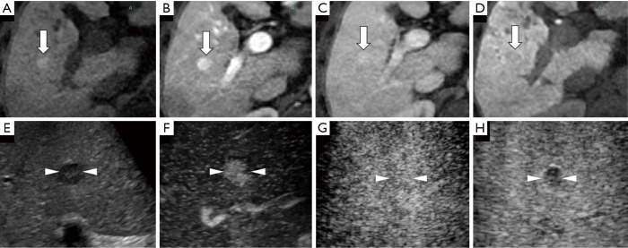
Figure 4.
A case of a well-differentiated hepatocellular carcinoma (HCC) lesion (maximum diameter, 15 mm) in segment V/VIII in a 56-year-old male patient with chronic hepatitis C-related disease. (A) Axial T1WI images on a plain scan show homogeneous hyperintensity in the tumor. The pre-contrast ratio was 1.199. (B,C) On the arterial phase (B, 25 seconds) and portal phase (C, 70 seconds) of gadolinium-ethoxybenzyl-diethylenetriamine pentaacetic acid magnetic resonance imaging (EOB-MRI), the lesion is shown as slightly hyperintense compared with surrounding background liver parenchyma. (D) Twenty minutes after EOB agent injection, the lesion shows as a homogeneous hypointensity (arrow) on the hepatobiliary phase. The post-contrast ratio was 0.616 and the EOB enhancement ratio was 0.514. (E) Grayscale US shows a homogeneous hypo-echoic lesion with oval shape and clear margin. No halo is seen around the lesion. (F) Using low mechanical index contrast imaging, the arterial phase of Sonazoid contrast-enhanced ultrasound (SCEUS) shows hypervascular enhancement. (G,H) The lesion shows isovascularity in the portal phase (G) and slightly hypoechoic (H) in the post-vascular phase (late washout). The arrows seen in (A,B,C,D) and the arrowheads seen in (E,F,G,H) indicate the margin of the lesion.

