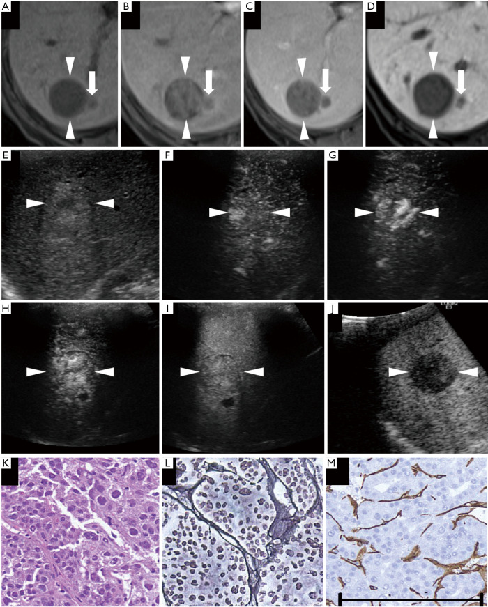
Figure 6.
A non-hypervascular moderately differentiated hepatocellular carcinoma (HCC) lesion (maximum diameter, 30 mm) in segment VI/VII was found in a 76-year-old female patient with chronic hepatitis C. (A) Axial T1WI images on a plain scan show homogeneous hypointensity in the tumor, with a regular oval shape and distinct margin. The pre-contrast ratio was 0.60. (B,C) On the arterial phase (B, 25 seconds) and portal phase (C, 70 seconds) of gadolinium-ethoxybenzyl-diethylenetriamine pentaacetic acid magnetic resonance imaging (EOB-MRI), the lesion is shown as a hypointensity compared with adjacent liver parenchyma. (D) Twenty minutes after EOB agent injection, the lesion was clearly shown as a homogeneous hypointensity on the hepatobiliary phase of EOB-MRI. The post-contrast ratio was 0.403 and the EOB enhancement ratio was 0.670. (E) On the grayscale ultrasound (US), the targeted lesion appears as an ill-defined, heterogeneous, hyperechoic lesion, with a small hypoechoic area in the center. (F,G,H) Using low mechanical index contrast imaging, the arterial phase of Sonazoid contrast-enhanced ultrasound (SCEUS) shows a significant hypervascular area with centripetal vessels. (I) During the portal phase, the lesion appears as a heterogeneous isovascularity. (J) The post-vascular phase image shows a hypoechoic lesion (late washout). (K) Hematoxylin-eosin (HE) staining shows obvious cancer cell atypia and hypercellularity. Cancer cells are arranged disorderly. (L) Silver staining shows that reticular fibers are sparsely distributed and not very clear. (M) Diffuse expression of cluster of differentiation 34 suggests increased neovascularization, resulting from sinusoidal capillarization and formation of sinusoidal vascular endothelium in HCC. The arrowheads seen in (A,B,C,D,E,F,G,H,I,J) indicate the margin of the lesion. Arrows in (A,B,C,D) depict a small satellite lesion beside the lesion. The black bar length in the lower side of (M) represents 200 microns (µm) as a magnification reference for (K,L,M). This lesion was pathologically diagnosed as a moderately differentiated HCC. (A,B,D,G,J) are quoted from (26).

