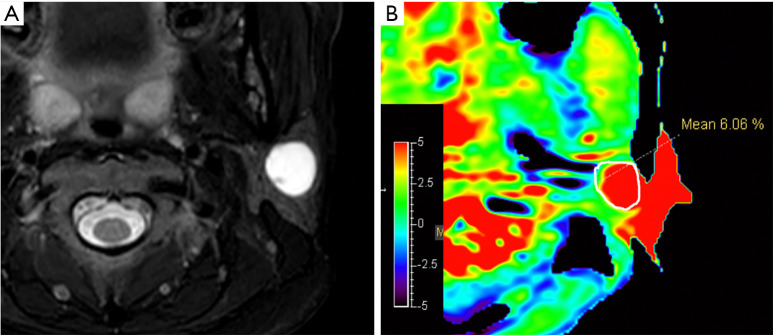
Figure 7.
An APTw image example of a hyperintensity artifact caused by cysts. A hyperintensity-signal lesion was found in the left parotid gland of 39-year-old female, as shown on T2WI (A). The pathological result was pleomorphic adenoma with cystic change. On the APTw (B) image, the integrity score was 4. The image quality in terms of hyperintensity artifacts scores was initially scored 4 by one radiologist and 2 by another. Eventually, the radiologists agreed on an image quality score of 2 because the hyperintensity in the cystic part of image was strongly affected by the artifacts diffused from the ear before the skull was hidden from the APT images.

