Figure.
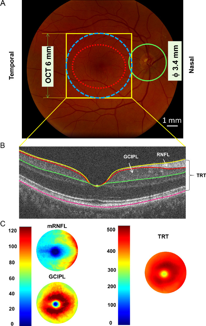
Retinal thickness measurements using optical coherence tomography. (A) UHR-OCT was used to scan the fovea with a 6 × 6 mm scan protocol (yellow rectangular area). The data were analyzed in the area of 6 mm disk (blue dotted circle) centered on the fovea to obtain tomographic thickness maps. In comparison, the Zeiss elliptical area (red dotted line) was also used to obtain the average thickness of ganglion cell-inner plexiform layer (GCIPL), imaged using Zeiss Cirrus OCT. In addition, the peripapillary retinal fiber layer (pRNFL, green circle) was also scanned using Zeiss Cirrus OCT. (B) Three intraretinal layers were segmented from the dataset acquired using UHR-OCT. These segmented layers included total retinal thickness (TRT), macular retinal never fiber layer (mRNFL), and ganglion cell-inner plexiform layer (GCIPL). (C) Intraretinal layer thickness was calculated using EDTRS partition, and annular thickness in the annulus from 1 to 6 mm in diameter was used. OCT, optical coherence tomography; UHR-OCT, ultra-high resolution optical coherence tomography, ETDRS, Early Treatment Diabetic Retinopathy Study.

