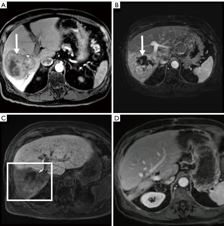
Figure 3.
Post contrast MRI demonstrating a right lobe hepatocellular carcinoma (arrow) with a small FLR (A). Post hepatocyte contrast administration MRI obtained 6 months after initial TARE demonstrates tumor necrosis (arrow) (B), interval FLR hypertrophy, and poor excretory function of the FRS (square) (C). Post contrast MRI image obtained 14 months after resection demonstrates no tumor recurrence or evidence of liver failure (D). The patient remains free of recurrence 30 months after resection. FLR, future liver remnant; TARE, transarterial radioembolization; FRS, future resection site.

