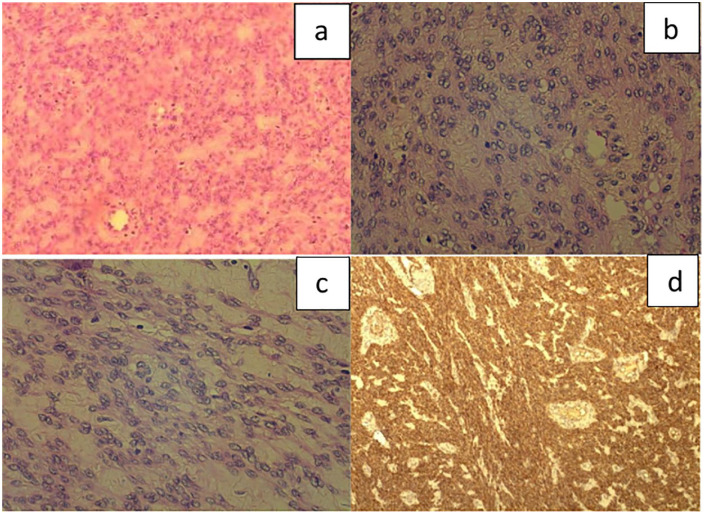
Figure 3.
Histological examination of section. Smooth muscle neoplasm identified with epithelioid morphology (a) (H–E ×100) and diffuse low to moderate nuclear atypia throughout the tumour (b) and (c), (H–E ×400). The mitotic count was low with less than five mitoses or 10 HPF. Areas of necrosis were not observed. Immunohistochemically, the neoplastic cells stained positive for calponin (d) (×40) and were negative for p16 and p53.

