Figure 2.
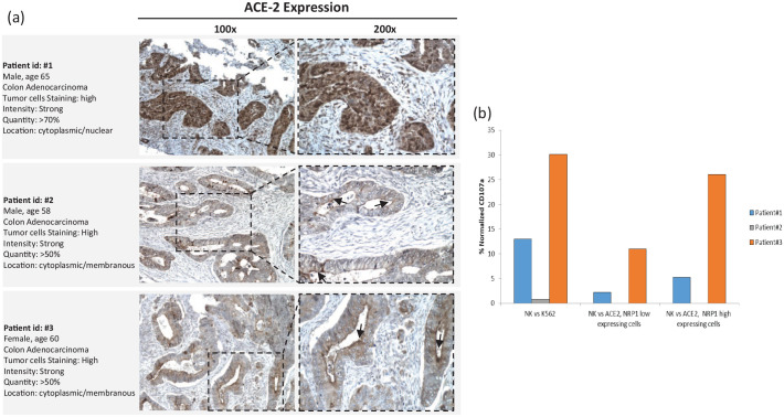
Representative whole section IHC staining of ACE-2, and patients’ peripheral NKs display higher degranulation toward ACE-2/NRP-1-expressing cells.
(a) Representative whole section IHC (immunohistochemistry) staining of ACE-2. Staining was done with HPA000288 Sigma-Aldrich, 1:250 dilution, HIER pH9. Patient 1 showed strong, diffuse cytoplasmic/nuclear staining for ACE-2 in colonic cancer cells. Patient 2 showed strong, diffuse cytoplasmic/membranous ACE-2 expression, with apical linear staining pattern, to seal the apical intercellular spaces of glandular epithelia and interfacing to the paracellular macromolecules (arrow). Patient 3 showed strong and diffuse cytoplasmic/membranous ACE-2 expression with apical linear staining pattern (arrow). (b) Patients peripheral NKs display higher degranulation toward ACE-2-/NRP-1-expressing cells. HCT 116, human colon cancer cells, were low ACE-2 and NRP-1 expressing; IGROV-1, human ovarian cancer cells, were high ACE-2 and NRP-1 expressing (Tumor cell lines were puchased from American Tissue Cultutre Collection, www.lgcstandards-atcc.org). Purified NK cells (0.1 × 10⁶) from patients’ peripheral blood were incubated with K562 cells (E:T ratio 10:1) and degranulation was evaluated through the lysosomal protein LAMP-1 (CD107a-PE, Clone H4A3, BD Pharmingen™). Patient 1 and patient 3 NKs displayed higher activity toward ACE-2-positive/NRP-1-positive-cells.
ACE-2, angiotensin-converting enzyme 2; E, effector; NK, natural killer cells; NRP, neuropilin; T, target.

