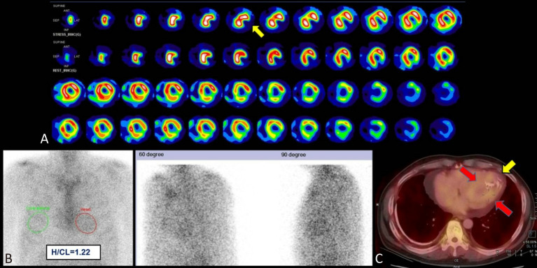
Figure 5.
AL amyloidosis. A 60-year-old man who presented with paroxysmal nocturnal dyspnea and orthopnea was diagnosed with heart failure with preserved ejection fraction of 58.2% (NYHA Functional Classification II). Standardized dipyridamole stress myocardial perfusion imaging showed scarring at the apical inferolateral wall (A, yellow arrow), but cardiac angiography revealed patent coronary arteries (figure not shown). Further 99mTc-PYP planar and SPECT/CT images demonstrated diffuse, mildly elevated tracer activity in the LV myocardium at 3 hours post-injection (B, red circle; and C, red arrows), equal to rib activity (visual score of 2). The H/CL ratio at 3 hours post-injection was 1.22. SPECT/CT images revealed the increased tracer uptake unseen at the LV apex, corresponding to an area of scarred myocardium (C, calcification seen at yellow arrow). Endomyocardial biopsy confirmed the patient has AL amyloidosis. Interstitial plasmacytosis (10-20%) was seen in the bone marrow biopsy, and a diagnosis of plasma cell myeloma was given. AL, immunoglobulin light chain amyloidosis; H/CL, heart to contralateral lung; LV, left ventricular; NYHA, New York Heart Association; PYP, pyrophosphate; SPECT/CT, single-photon emission computed tomography/computed tomography.

