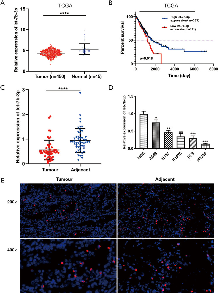
Figure 1.
Let-7b-3p expression was decreased in LUAD tissues and cell lines. (A) Let-7b-3p expression in TCGA database. (B) Kaplan-Meier analysis of overall survival according to TCGA database. (C) Let-7b-3p expression in clinical specimens was detected by qRT-PCR (n=50). (D) Let-7b-3p expression in cell lines was assessed by qRT-PCR. (E) FISH of LUAD tumor tissues and corresponding adjacent nontumorous tissues. The probes were labeled by Cy3 (red) and the nuclei are counter-stained with DAPI (blue) (*, P<0.05; **, P<0.01; ***, P<0.001; ****, P<0.0001). LUAD, lung adenocarcinoma; TCGA, The Cancer Genome Atlas; qRT-PCR, quantitative real-time polymerase chain reaction; FISH, fluorescence in situ hybridization.

