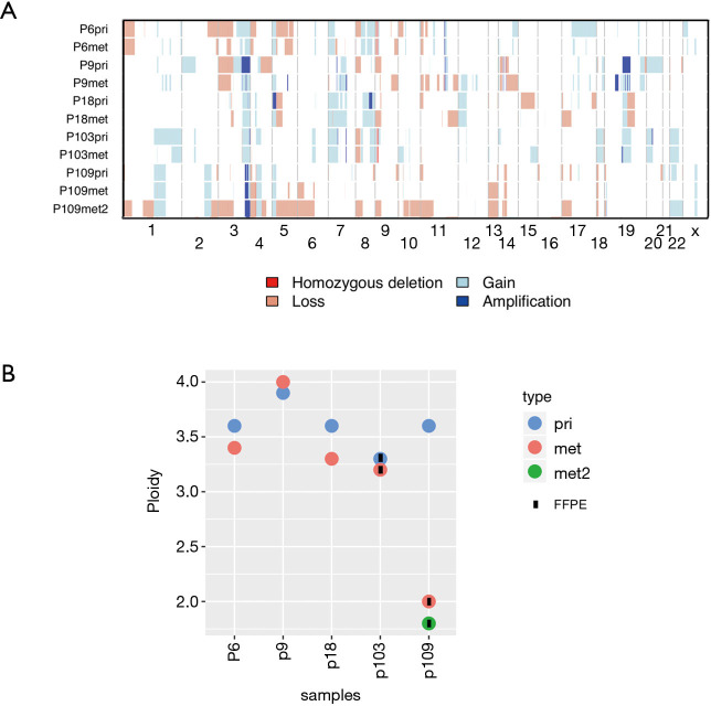
Figure 3.
Similarity of primary tumors and metastases on chromosomal level. (A) Repertoire of copy number alterations as defined by WES of primary LUSC (pri), first metastasis (met) and second metastasis (met2) of each patient. Samples represented on the y-axis; chromosomes are represented along the x-axis. Light red: copy number loss; red: homozygous deletion; light blue: copy number gain; dark blue: amplification; (B) ploidy analysis measured using flow cytometry. Samples are represented on the x-axis; ploidy is represented in the y-axis. blue: primary tumor (pri); red: first metastasis (met); green: second metastasis (met2).

