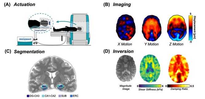
Figure 1.

Overview of the high-resolution, HCsf-specific MRE protocol. (A) Vibrations are delivered from an active driver to a head pillow-driver to generate harmonic shear waves; (B) shear waves are imaged using a high-resolution MRE sequence; (C) HCsf regions are segmented using Automated Segmentation of Hippocampal Subfields (ASHS) into the dentate gyrus/cornu ammonis 3 (DG-CA3), cornu ammonis 1–2 (CA1-CA2), subiculum (SUB), and the entorhinal cortex (ERC); (D) the nonlinear inversion algorithm (NLI) calculates the shear stiffness (μ) and damping ratio (ξ) property maps for each HCsf region from displacement data using the HCsf segmentations as spatial priors via soft prior regularization (SPR).
