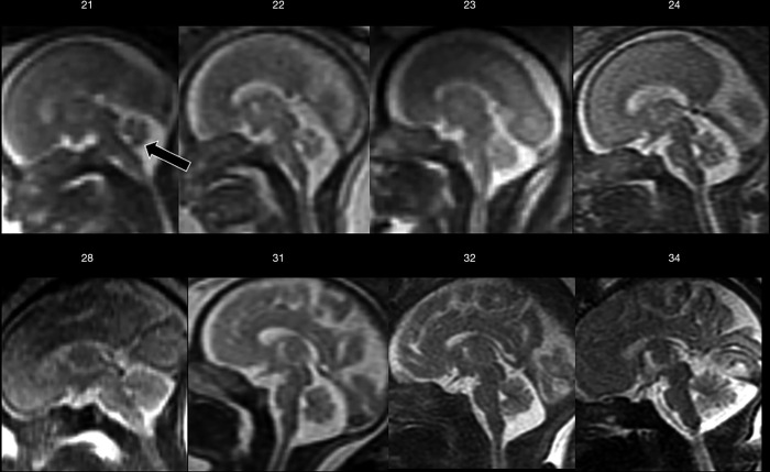
Figure 5.
Mid-sagittal T2-weighted ss-FSE sections from increasing gestational age (week indicated) showing progressive sulcation and the relationship between cerebral hemispheres and cerebellar vermis through gestation. Note how already at 21-week gestation the vermis completely covers the fourth ventricle (arrow). ss-FSE, single-shot fast spin echo.

