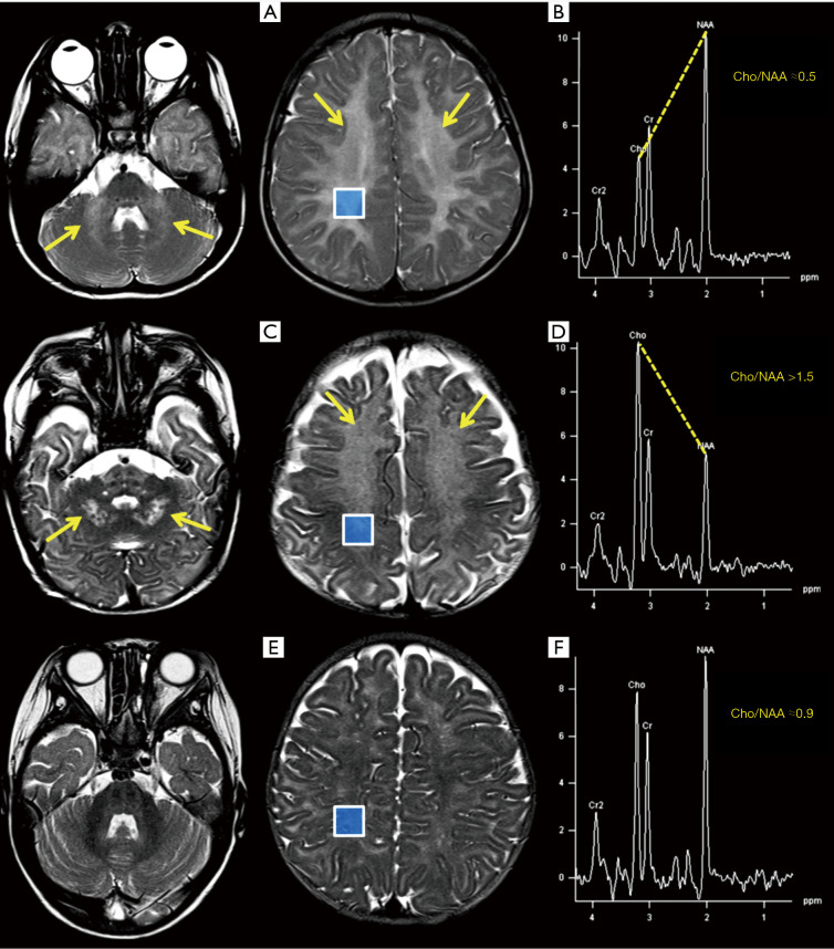
Figure 14.
Top row: 4-year-old boy with ataxia as the main symptom, suffering from “4H syndrome” (hypomyelinating leukodystrophy) suspected by imaging first, with a confirmed novel mutation of POLR3A gene. MRI axial T2-WI (A): diffuse pathologic WM hyperintensities are evident (arrows pointing at middle cerebellar peduncles and centrum semiovale). The intermediate TE spectrum in the right parietal WM confirms that tCho/NAA (≈0.5) and other metabolites ratios are within normal ranges for this age (B). Middle row: 5-month-old male with severe hypotonia and confirmed Krabbe disease (demyelinating leukodystrophy) showing pathological T2 hyperintensities in deep cerebellar WM and centrum semiovale with a typical “leopard skin” pattern (arrows in C). The spectrum in (D), obtained from the same area as those in (B) and (F), shows a tCho/NAA ratio > 1.5, abnormal even at this age. Bottom row: a 5-month-old female imaged for cryptogenic epilepsy is shown for comparison with case in (C): normal myelination (E) and metabolites ratios (tCho/NAA ≈0.9) in parietal WM at this age (F). Acronyms and abbreviations are shown in Appendix 1.

