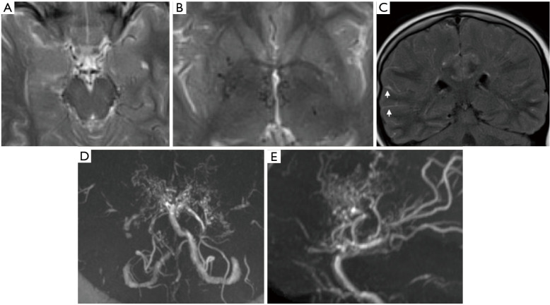
Figure 5.
Moyamoya syndrome in a 14 years old girl. Axial T2WI (A and B) demonstrate prominence of flow voids in the suprasellar cistern with significant narrowing of the ICA bilaterally. There are also multiple perforators noted in the basal ganglia and thalami. Coronal FLAIR image (C) shows the characteristic ‘ivy sign’ (arrows). TOF MR angiogram (D and E: MIP images) demonstrates bilateral distal carotid and A1 stenosis, as well as reduced M1 flow. There are numerous leptomeningeal collaterals. TOF, time of flight.

