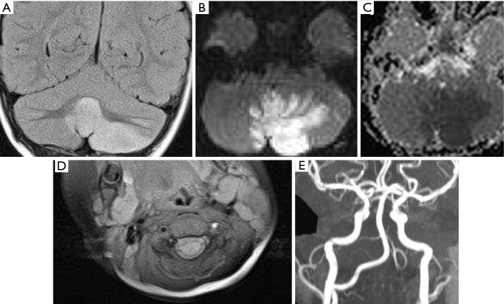
Figure 6.
Vertebral artery dissection. Coronal FLAIR image (A) shows signal abnormality within the left cerebellar hemisphere and cerebellar vermis. Diffusion weighted imaging (B: B1000 and C: ADC) show diffusion restriction corresponding to the FLAIR signal abnormality in keeping with acute infarction. Axial T1W fat saturated image (D) shows intrinsic T1W hyperintensity in the upper cervical segment of the left vertebral artery. Time-of-flight MRA (E) shows a corresponding lack of normal flow within the left vertebral artery related to dissection. FLAIR, fluid attenuated inversion recovery; ADC, apparent diffusion co-efficient; MRA, MR angiogram.

