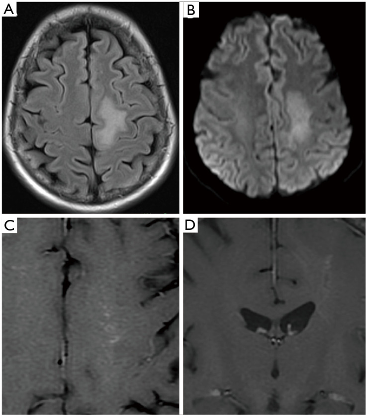
Figure 7.

Biopsy proven vasculitis in a 13-year-old child who presented with history of worsening gait and left sided neck pain. Axial T2WI (A) and axial DWI B1000 (B) images demonstrate a hazy signal abnormality within the left centrum semiovale with corresponding mild diffusion restriction. Post contrast axial and coronal T1WI (C and D) show patchy serpiginous enhancement within this region. DWI, diffusion weighted imaging.
