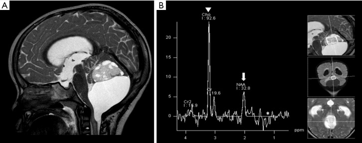
Figure 3.
Solid cystic mass in the posterior fossa was confirmed as pilocytic astrocytoma. Brain MRI: Sagittal T2WI, and 1H MRS (A,B). Reduced NAA (arrow), increased choline (arrowhead), and possible mild lactate (asterisk) peaks were observed (B). T2WI, T2-weighted image; MRS, magnetic resonance spectroscopy; NAA, N-acetylaspartate.

