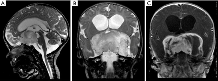
Figure 4.
Male, 2 years old, diagnosis of pilomyxoid astrocytoma. Brain MRI: Sagittal T2WI, coronal T2WI and T1W post-contrast (A,B,C) show a suprasellar infiltrative mass hyperintense on T2WI, with cystic components peripherally distributed, and heterogeneous post-contrast T1WI enhancement (C), which extends into the temporal lobes, giving an “H shape” on coronal imaging (B,C). T1W, T1-weighted; T2WI, T2-weighted image.

