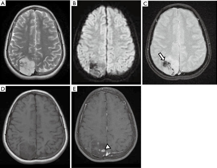
Figure 9.
Female, 8 years old, diagnosis of DNET. Brain MRI: T2WI, DWI, SWI, T1WI, and T1W postcontrast (A,B,C,D,E) show a predominantly cortical lesion in the right occipital lobe hyperintense on T2WI, with a ‘bubbly appearance,’ and remodeling the adjacent inner table of the skull vault (A). There were no areas of restriction on DWI (B). Low-signal foci were observed on SWI, confirmed as components of calcium on a CT scan not shown (arrow, C). Some small areas of enhancement were noted (arrowhead, E). DNET, dysembryoplastic neuroepithelial tumor; DWI, diffusion weighted imaging; SWI, susceptibility weighted imaging; T1WI, T1-weighted image; T2WI, T2-weighted image.

