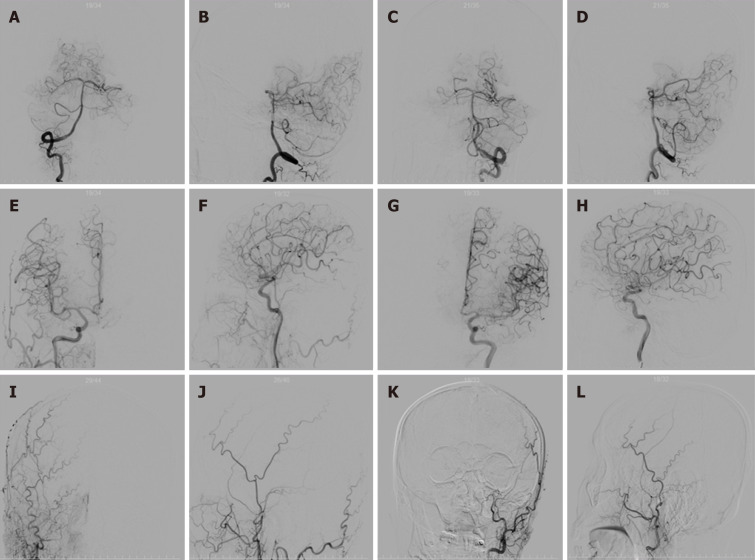
Figure 2.
Cerebral angiography on the day of admission. A: Right vertebral artery angiography; B: Lateral angiography of the right vertebral artery; C: Anterior angiography of the left vertebral artery; D: Lateral angiography of the left vertebral artery; E: Right internal carotid artery angiography; F: Lateral angiography of the right internal carotid artery; G: Angiography of the left internal carotid artery; H: Lateral angiography of the left internal carotid artery; I: Right external carotid artery angiography anteroposterior; J: Lateral angiography of the right external carotid artery; K: External left carotid artery angiography anteroposterior; L: Lateral angiography of the left external carotid artery.

