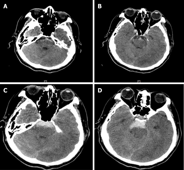
Figure 3.
After lumbar puncture, blood was dissolved. Four days after the initial bleeding, non-contrast computed tomography (CT) showed the typical pattern of perimesencephalic spontaneous subarachnoid hemorrhage (SAH). A: After lumbar puncture, blood was dissolved; B: CT shows blood was dissolved; C: The typical pattern of perimesencephalic SAH; D: The typical pattern of perimesencephalic SAH.

