Figure 5.
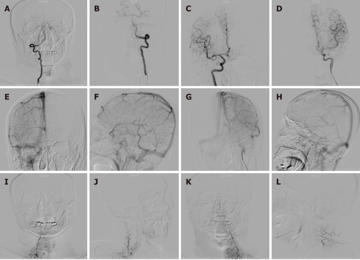
Six-vessel cerebral catheter angiograms of bilateral thyrocervical and costocervical trunks ruling them out as sources of spontaneous subarachnoid hemorrhage. A and B: Bilateral vertebral artery injection [arterial phase; anteroposterior (AP) projection]; C and D: Bilateral internal carotid artery injection (arterial phase; AP projection); E and F: Right internal carotid artery injection (venous phase; AP and lateral projections); G and H: Left internal carotid artery injection (venous phase; AP and lateral projections); I and J: Right thyrocervical trunk injection (arterial phase; AP and lateral projections); K and L: Left thyrocervical trunk injection (arterial phase; AP and lateral projections). Internal carotid artery angiography illustrated the drainage of basal vein of Rosenthal (BVR), which does not drain into the vein of Galen due to discontinuous BVR (E-H).

