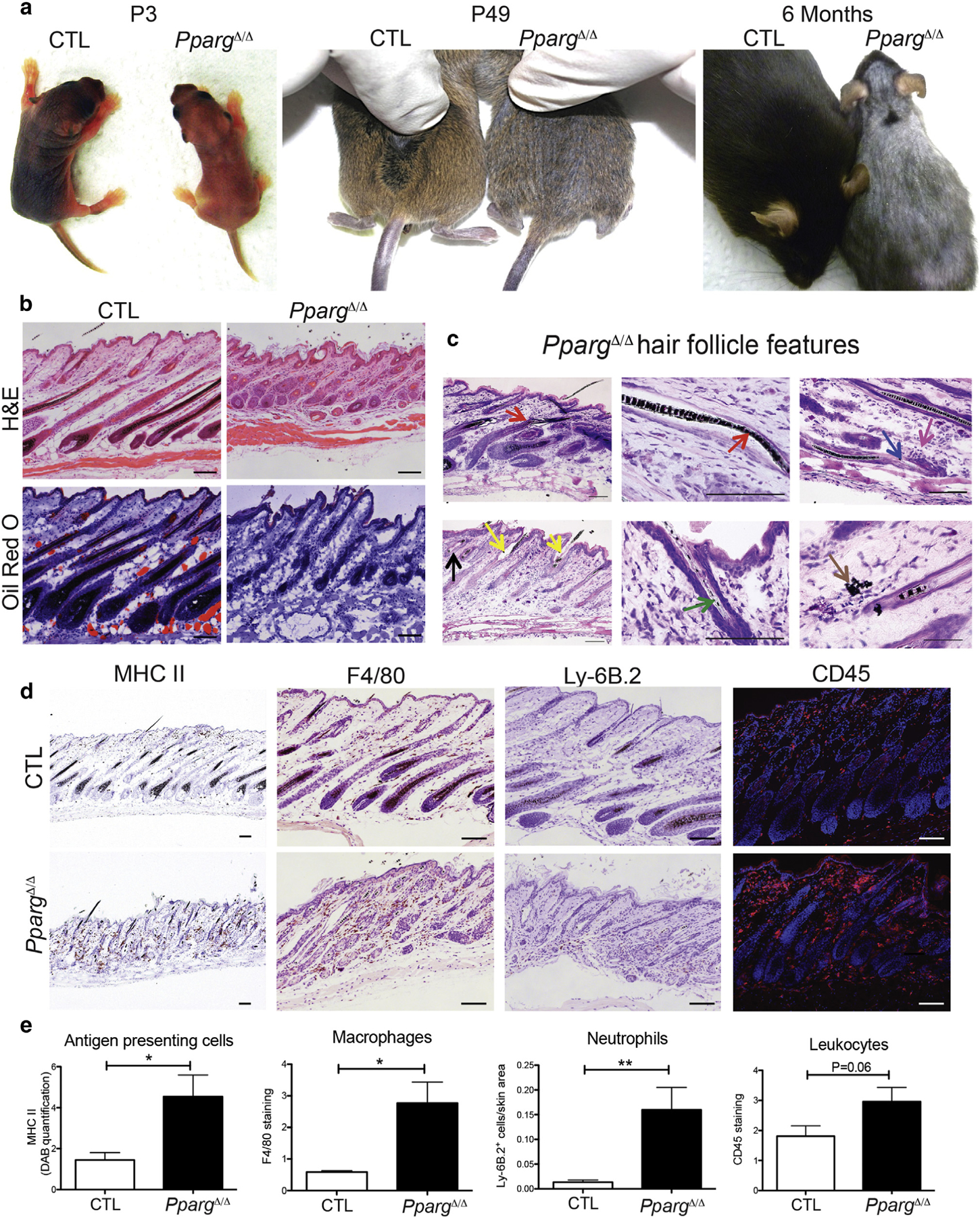
Figure 1. Spontaneous skin and hair phenotype in PpargΔ/Δ mice.

(a) Pictures of Sox2-Cretg/+PpargΔ/emΔ (PpargΔ/Δ) and control (CTL) mice at P3, P49, and 6 months. (b) Hematoxylin and eosin and Oil Red O staining of back skin sections from PpargΔ/Δ and CTL mice at P28. (c) Giemsa-staining showing dystrophic features of the HF (arrows) at P28: follicular plugging (black), perifollicular inflammation (fuchsia), HF disruption (blue), or deformation (red) with widened hair canal (yellow), irregular melanin banding pattern (green), and extrafollicular deposits of melanin (brown). (d) Immunostaining with anti-MHC II (activated phagocytic cells), anti-F4/80 (macrophages), anti-Ly-6B.2 (neutrophils), and anti-CD45 (leukocytes). (e) Quantification of the stainings in d. Data expressed as mean ± standard error of the mean (n = 3). Scale bars =100 μm. CTL, control; H&E, hematoxylin and eosin; MHC, major histocompatibility complex; P, postnatal day.
