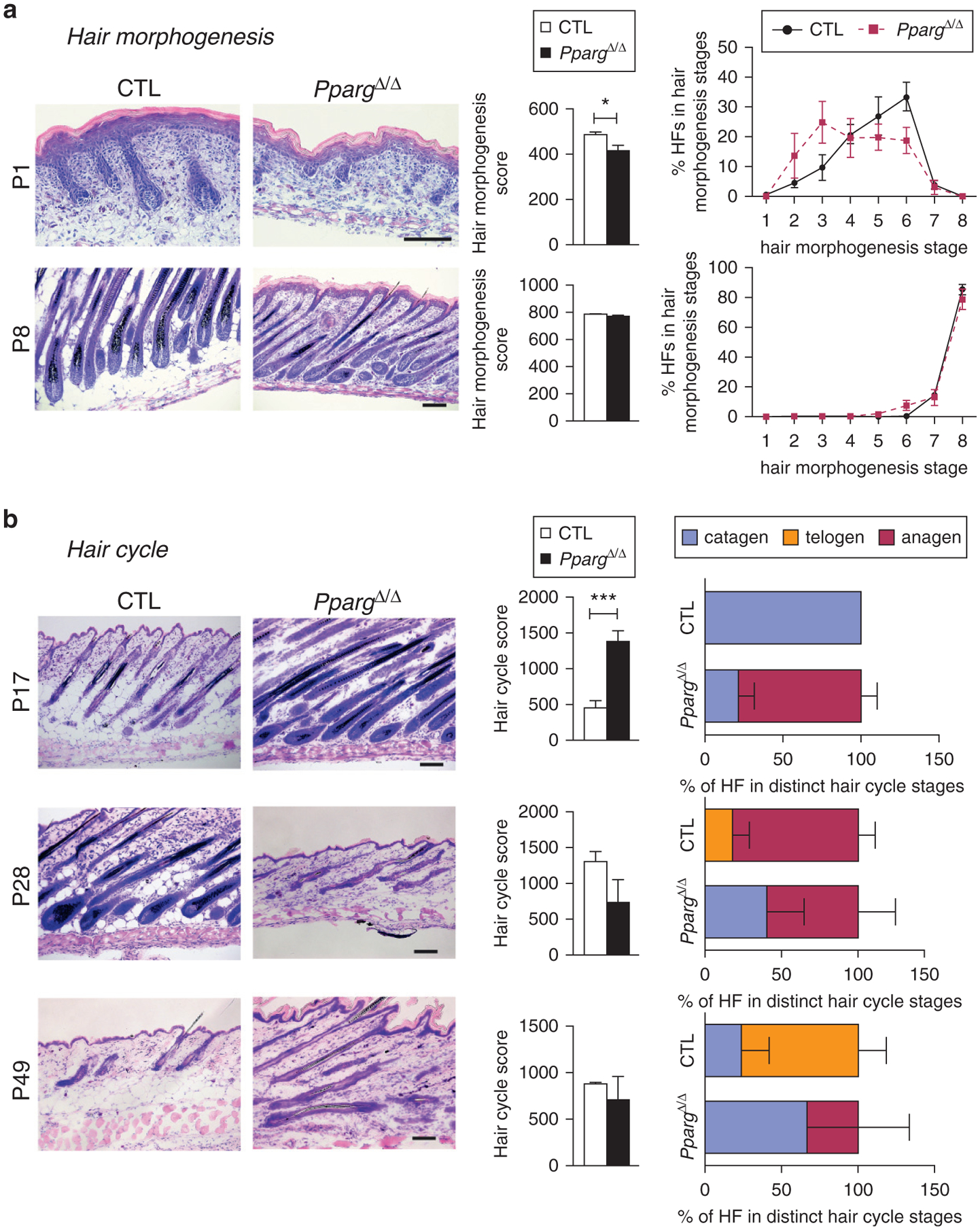
Figure 2. Delayed hair follicle morphogenesis and abnormal hair follicle cycling in PpargΔ/Δ mice.

Quantitative histomorphometry of Giemsa-stained back skin cryosections from Sox2-Cretg/+PpargΔ/emΔ (PpargΔ/Δ) and control (CTL) mice collected at the indicated postnatal days. (a) Hair morphogenesis analysis at P1 and P8: representative pictures (left panel, scale bar = 100 μm), hair morphogenesis score (central panel), and percentage of HFs found in the different hair morphogenesis stages (right panel, n = 6). (b) Hair cycle progression analysis at P17 (catagen), P28 (anagen), and P49 (anagen): representative pictures (left panel, scale bar = 100 μm); hair cycle score (central panel); percentage of HFs found in the different hair cycle stage (right panel; n = 4–5). Score values are expressed as mean ± standard error of the mean. *P < 0.05 and ***P < 0.001, respectively. CTL, control; HF, hair follicle; P, postnatal day.
