FIGURE 2.

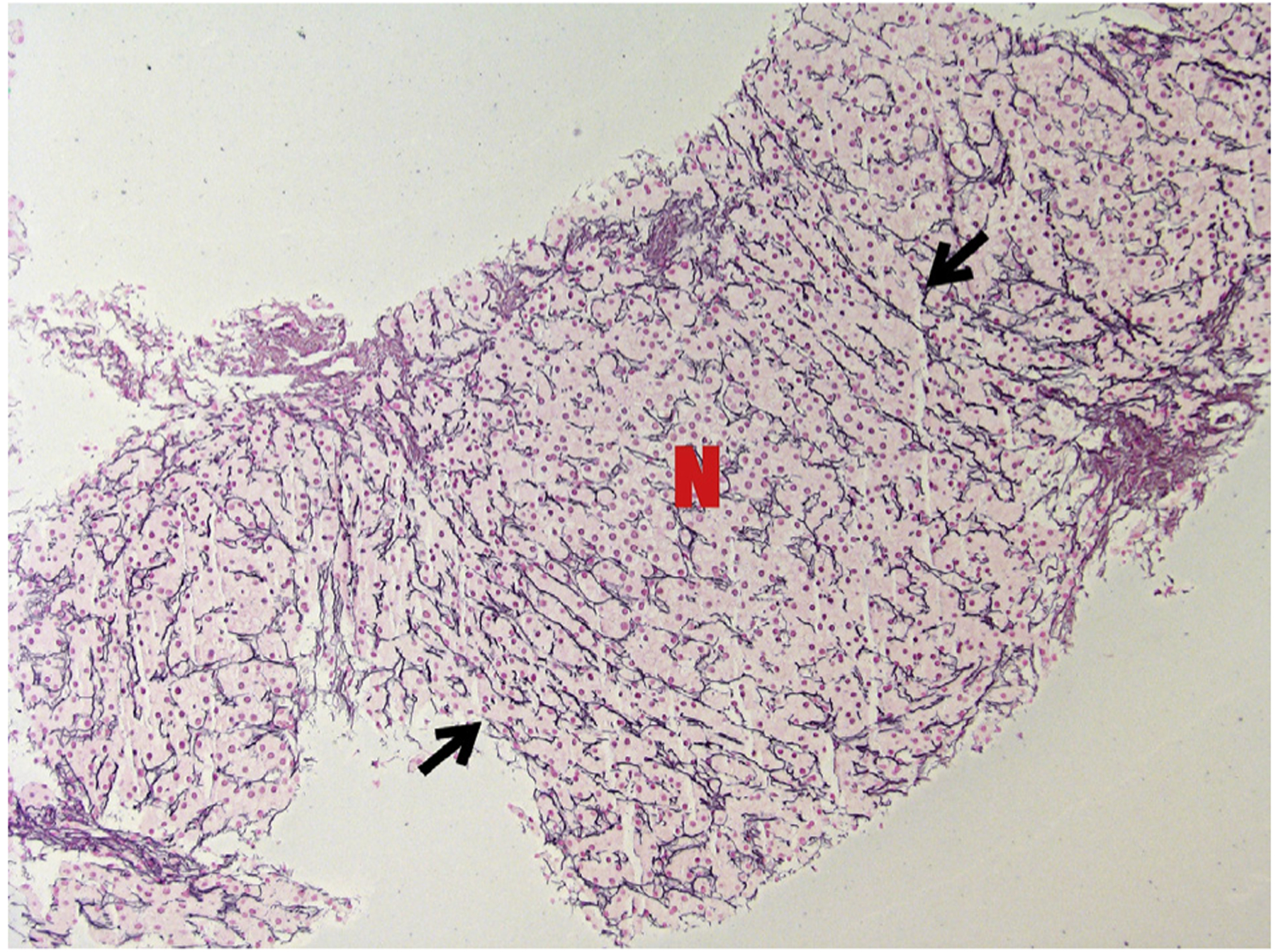
Reticulin stain of PI-3K patient’s liver biopsy showing a nodule where hepatocytes are arranged in thick plates (marked with N). At the borders of the nodule, compressed plates of hepatocytes marked with black arrows (magnification × 100).

Reticulin stain of PI-3K patient’s liver biopsy showing a nodule where hepatocytes are arranged in thick plates (marked with N). At the borders of the nodule, compressed plates of hepatocytes marked with black arrows (magnification × 100).