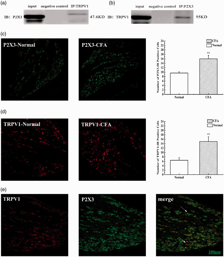
Figure 5.
TRPV1 is physically associated with P2X3 in CFA rats. (a and b) Representative co-immunoprecipitation bands between P2X3 and TRPV1 in L4-6 DRG of CFA rats. Negative control: IgG control. (c) The number of P2X3-IR positive cells in L4-6 DRG of CFA rats. (d) The number of TRPV1-IR positive cells in L4-6 DRG of CFA rats. (e) Immunofluorescence confocal micrographs of P2X3, TRPV1 and merge in L4–6 DRG of CFA rats. Sections show immunohistochemical red labeling for TRPV1 positive neurons, green labeling for P2X3 positive neurons. Scale bars=100μm.

