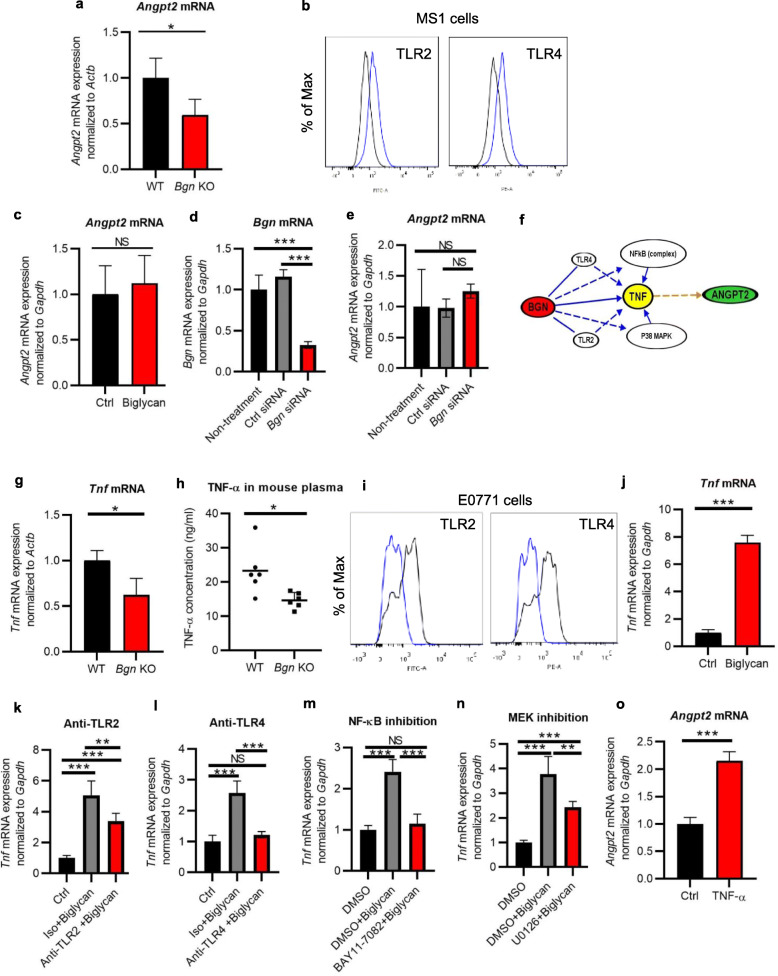
Fig. 3.
TNF-ɑ-enhanced Angpt2 expression is controlled by biglycan in E0771 tumors. a Angpt2 mRNA expression in E0771 tumor tissues of WT and Bgn KO mice by quantitative real-time PCR. b TLR2 and TLR4 expression in MS1 cells analyzed by FACS analysis. Blue: antibody. Black: isotype. c Angpt2 mRNA expression in MS1 cells treated with recombinant biglycan (5 g/ml) by quantitative real-time RT-PCR. d Bgn mRNA expression in E0771-TECs transfected with Bgn siRNA by quantitative real-time PCR. e Angpt2 mRNA expression in E0771-TECs transfected with Bgn siRNA by quantitative real-time PCR. f Flowchart of biglycan, TNF-ɑ, and Angpt2 signaling by IPA analysis. g Tnf mRNA expression by quantitative RT-PCR in E0771 tumor tissues from WT and Bgn KO mice. h TNF-ɑ secretion in the plasma of E0771 tumor-bearing mice was detected by ELISA. Each dot represents one mouse. i TLR2 and TLR4 expression in E0771 cells analyzed by flow cytometry. Blue: isotype. Black: antibody. j Tnf mRNA expression in E0771 cells treated with recombinant biglycan (5 μg/ml). k, l After blocking of TLR2 or (k) TLR4 (l), E0771 cells were stimulated by recombinant biglycan (5 μg/ml). Tnf mRNA expression was analyzed by quantitative RT-PCR. m, n After treating with (m) the NF-κB inhibitor (10 μM) or (n) the MEK inhibitor (20 μM), E0771 cells were stimulated by recombinant biglycan (5 μg/ml). Tnf mRNA expression was analyzed by quantitative real-time RT-PCR. o Angpt2 mRNA expression in MS1 cells treated with recombinant TNF-ɑ (10 ng/ml) by quantitative RT-PCR. *p < 0.05, **p < 0.01, ***p < 0.001. NS, not significant. Significance was determined in a, c, g, j, and o by two-tailed Student’s t test, and in d, e, k, l, m, and n by a one-way ANOVA. All data represent means ± SD

