FIGURE 1:
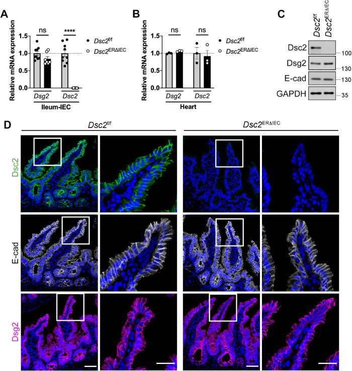
Loss of Dsc2 in IECs did not alter intestinal mucosal architecture as well as Dsg2 and E-cadherin expression. Intestinal epithelial-specific deletion of Dsc-2 was confirmed in the ileum of Dsc2ERΔIEC mice and control littermates Dsc2f/f treated with tamoxifen and analyzed 30 d later. (A) Loss of Dsc2 mRNA expression, but not of Dsg2 mRNA in ileal IECs isolated from Dsc2ERΔIEC mice or control Dsc2f/f. Points represent values from an individual mouse. Graph combines values obtained from two independent experiments with a total of seven to nine mice per group. Data are means ± SEM. ****p < 0.0001; significance is determined by two-tailed Student’s t test. (B) Expression of Dsc2 and Dsg2 mRNA is unchanged in the heart that does not express Villin, confirming tissue-specificity of Dsc2 depletion. Results are representative of two independent experiments. Points represent values from an individual mouse. Differences are not significant (ns) by two-tailed Student’s t test. (C) IECs were isolated from the ileum of Dsc2ERΔIEC and Dsc2f/f mice. Protein were separated by SDS–PAGE and visualized by immunoblotting with antibodies against Dsg2, Dsc2, E-cadherin, and GAPDH as loading control. Representative Western blot images showing loss of Dsc2 in the ileal epithelium in Dsc2ERΔIEC mice while Dsg2 and E-cadherin expression is unaltered. (D) Representative images of ileal tissue sections from tamoxifen-treated Dsc2ERΔIEC and Dsc2f/f mice stained with anti-Dsc2, anti-Dsg2 or anti-E-cadherin antibodies and DAPI as a nuclear counterstain. Dsc2 expression is absent in ileal crypt-villus epithelial cells of Dsc2ERΔIEC mice with no change in Dsg2 and E-cadherin expression. Scale bars are 50 μm.

