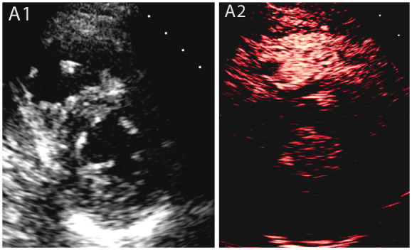
Figure 1.

Case 1: A male coronavirus disease 2019 patient who had a previous pulmonary embolism with a suspected right ventricular thrombus in the non-enhanced study, which was subsequently confirmed by contrast echocardiography. (A1) Parasternal short axis view shows a small right ventricular mass, (A2) contrast echocardiography of the same view, showing a non-perfused right ventricular mass suggestive of thrombus.
