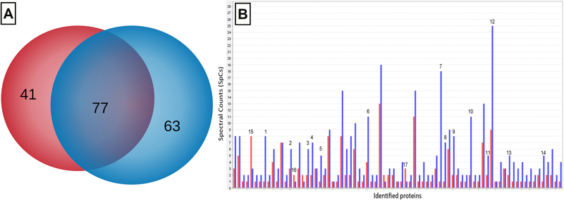
Fig. 3.
Intracellular proteomic analysis of F. tricinctum M6. Venn diagram of proteins identified in the presence (blue) and in absence (red) of Cu(II) in the culture medium; the group of proteins shared by both conditions is shown in violet (A). Bar diagram of the semi-quantitative expression of intracellular proteins detected in the presence (blue) and in absence (red) of Cu(II) in the culture medium (B).

