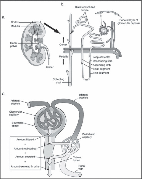
Figure 3.

Normal physiology of the kidney. (a) Vertical section of right kidney showing cortex and medulla. Inset (b) diagrammatic representation of a nephron within the cortex and medulla. (c) Diagrammatic representation of renal circulation showing dual capillary beds allowing filtration and reabsorption.
