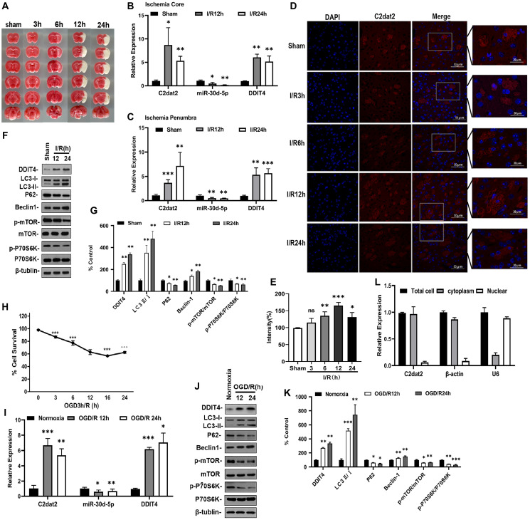
Figure 2.
lncRNA C2dat2, miR-30d-5p, and DDIT4 were involved in the response to I/R-induced injury in vivo and in vitro. (A) Representative images of TTC-stained brain sections. Representative images in mice after 1 h MCAO and 3, 6, 12, 24, and 36 h reperfusion. (B–C) C2dat2, miR-30d-5p, and DDIT4 levels in the ischemic core (B) and penumbra (C) of ischemic and sham tissue were measured via RT-qPCR. GAPDH was used as the control (n = 3 per group). (D) Representative RNA-FISH images manifesting intracellular localization. (E) Intensity of C2dat2 in each group. (F) Western blotting showing the expression levels of DDIT4 and autophagy-related protein expression levels (LC3, P62, Beclin-1, p-mTOR, mTOR, p-P70S6K, and P70S6K) in the ischemia penumbra of mice after 1 h MCAO and 12 and 24 h reperfusion. (G) Relative protein levels were analyzed. Data are mean ± standard error of the mean (SEM; n = 3). (H) OGD/R-induced cell death in N2a cells. (I) RT-qPCR of C2dat2, DDIT4, and miR-30d-5p levels in OGD-treated cells and normoxia control. (J) Typical Western blotting results showed changes in DDIT4 and autophagy-related protein (LC3, P62, Beclin-1, p-mTOR, mTOR, p-P70S6K, and P70S6K) expression levels in N2a cells between normoxia and 12 and 24 h reoxygenation after 3 h OGD. (K) Relative protein levels were analyzed. Data are mean ± SEM (n = 3). β-Tubulin was blotted as a loading control. *P < 0.05; **P < 0.01; ***P < 0.001; ns, not significant versus normoxia or sham. (L) Distribution of lncRNA C2dat2 in the cytoplasm and nucleus of N2a cells 24 h after OGD/R. Cell fractionation of U6, β-actin, and C2dat2 in N2a cells. Like β-actin, C2dat2 is expressed in the cytoplasm.

