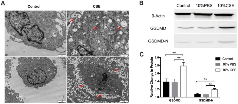
Figure 1.
CSE treatment induced pyroptosis in HAECs. (A) Morphology of HAECs treated with and without CSE imaged using electron microscopy. Autophagosomes, cytoplasmic outflow and cell membrane break indicated by red arrows. (B, C) The protein levels of GSDMD and GSDMD-N were upregulated in HAECs after treatment with CSE, as indicated by western blot results. β-Actin was used as an internal control. **p < 0.01. The data are represented as mean ± SD (n = 3).

