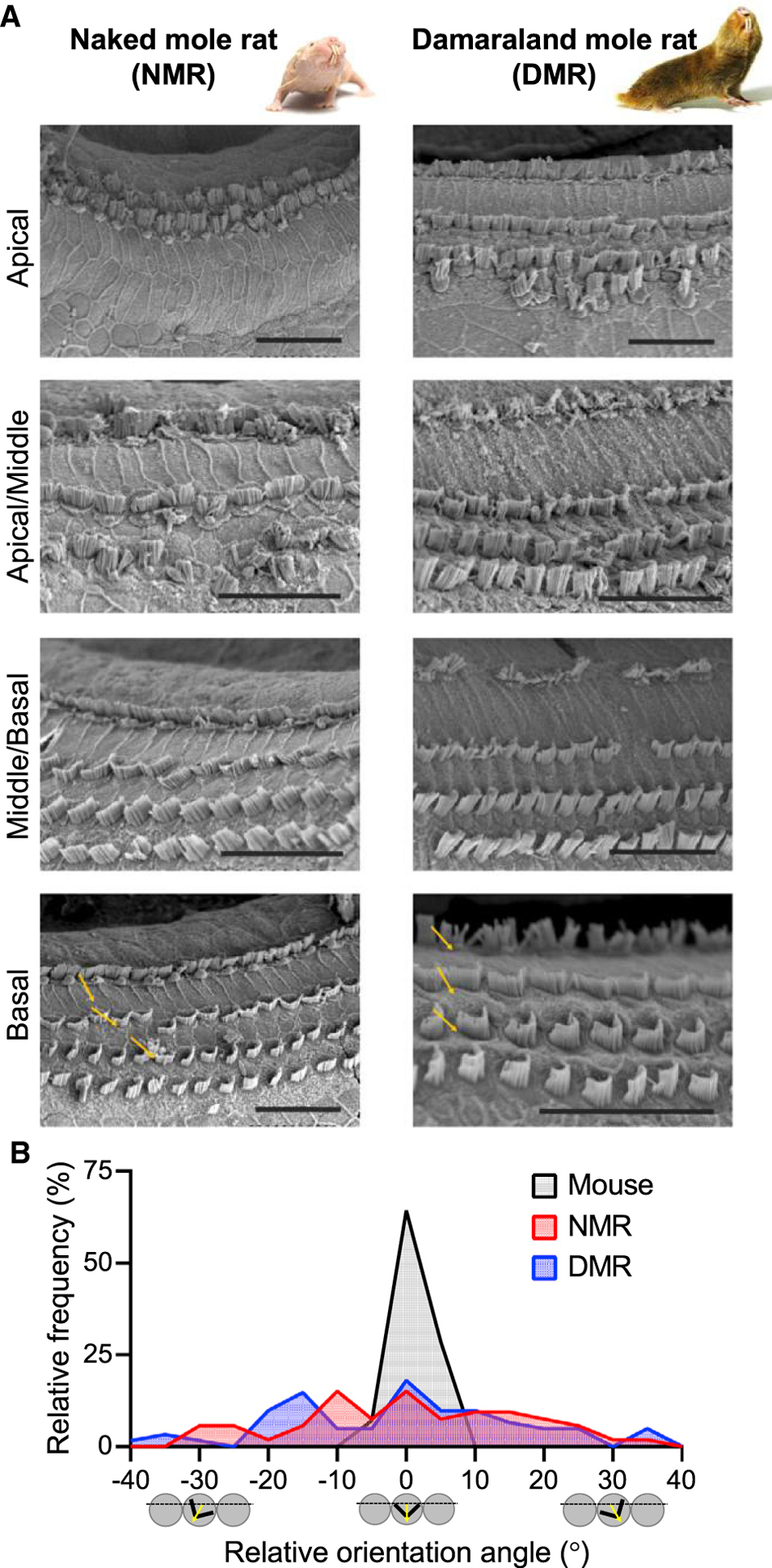
Figure 3. Hair Bundle Morphology and Orientation Is Disrupted in Bathyergid Mole-Rats.

(A) Scanning electron micrographs show absent and/or disorganized OHC hair bundles in apical cochlear regions in both naked and Damaraland mole-rats. Hair bundle morphology improves in progressively more basal cochlear regions but nevertheless shows considerable variation in orientation (yellow arrows). Scale bars, 25 μm.
(B) OHC hair bundle orientation angles from basal cochlear regions show broader distributions in both naked mole-rats (red) and Damaraland mole-rats (blue) compared with mice (black; shown here for comparison).
