
Fig. 5.
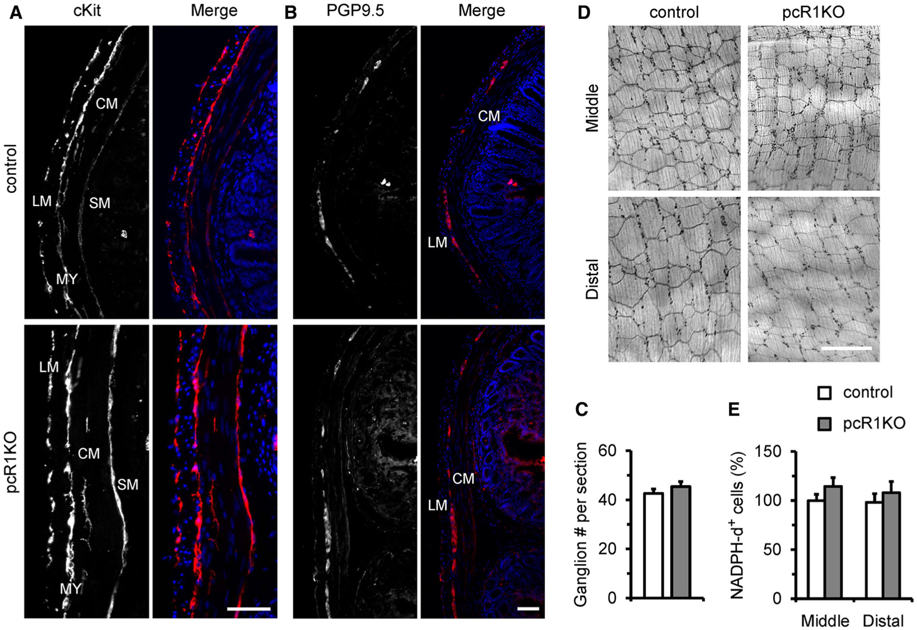
Distribution of ICCs and enteric neurons were not altered in pcR1KO colons. a Representative transverse sections of control and pcR1KO colons labeled with cKit to identify ICC distribution. In both control and pcR1KO sections, cKit-positive cells could be found in the longitudinal muscle (LM) layer, circular muscle (CM) layer, myenteric plexus (MY), and submucosal surface (SM). Data are representative of at least three independent experiments. Scale bar, 50 μm. b Representative transverse sections of control and pcR1KO colons labeled with the neuronal marker, PGP9.5. PGP9.5-positive cells were observed in the myenteric plexus between the longitudinal (LM) and circular (CM) muscle layers. Data are representative of at least three independent experiments. Scale bar, 50 μm. c The number of ganglions per section were calculated in control and pcR1KO mice. At least three independent sections were used for each mouse. n = 5 mice per group. d Whole-mount staining for NADPH diaphorase showing the distribution of nNOS-positive inhibitory neurons in middle and distal colons from control and pcR1KO mice. e Normalization of the densities of NADPH diaphorase-positive (NADPH-d+) cells in control and pcR1KO colons. Scale bar, 400 μm; n = 4 mice per group. Significance was determined using a 2-tailed, unpaired Student’s t test. Error bars represent mean ± SEM