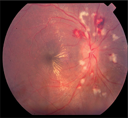
Figure 1.

Retina, right eye. Blurring of the optic disc margin and extensive cotton‐wool spots, or nerve fiber layer infarcts, are seen surrounding the optic nerve. Flame‐shaped hemorrhages, diffuse exudates, and a hemimacular star are also present. An exudative retinal detachment was seen on clinical examination extending through the fovea. (Note: This cannot be appreciated on the two‐dimensional view.)
