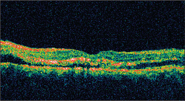
Figure 4.

Optical coherence tomography, left eye. Area of reflectivity (dark area) in the subretinal space is consistent with subretinal fluid and an exudative retinal detachment. The areas of hyperreflectance (red and orange) in the middle retinal areas are consistent with hard exudates. These areas show localization of the hard exudates in the outer plexiform layer (or Henle's layer) of the retina.
