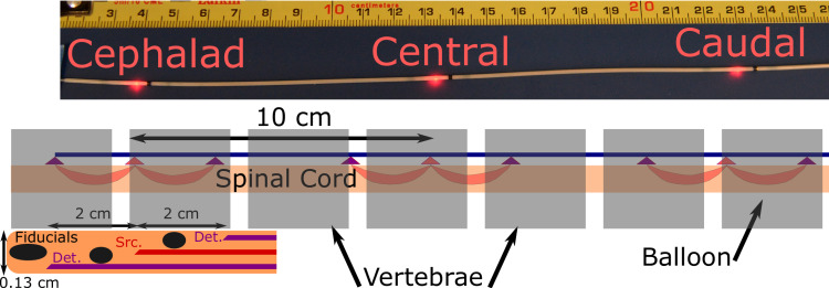
Fig 1. Photo of 3 position probe, all three source fibers illuminated.
Black regions of probe are combined radiographic fiducials and blocks to prevent light propagation down the length of the probe. Schematic of probe in spine and expanded view of probe distal tip, showing fiducials at tip and between each source-detector pair, as well as the fiber positions. Each of the three detector-source-detector combinations along the probe is configured in a similar fashion. Not to scale.

