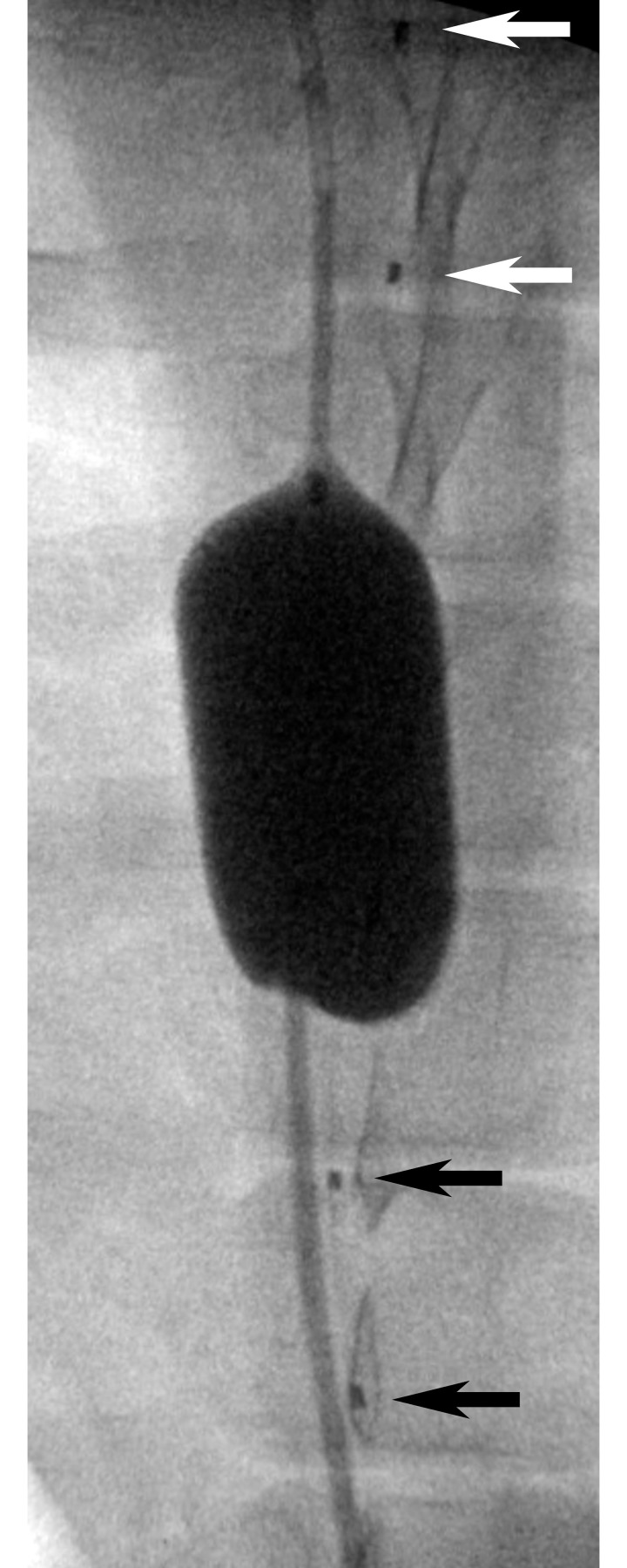
Fig 3. Anterior-posterior radiograph of an REOBA balloon.
The balloon was inflated in the aorta between the caudal (black arrows) and central (white arrows) positions of a FLOXsp probe placed on the along the spinal cord in the epidural space. Fiducial markers noted by arrows are located between each light source and corresponding pair of detector positions (Fig 1).

